Diagnosis
Nerve conduction study and EMG: what to expect
Two tests, usually done in one sitting. One sends small electrical pulses along a nerve and times them. The other listens to a muscle through a fine needle. Here is what they measure, how they feel, and what a normal result does and does not rule out.
Written by the Managing Neuropathy editorial team under the direction of Dr. Tom Biernacki, DPM, FACFAS · Clinical review pending · Last updated 24 September 2026
The short answer. A nerve conduction study measures how fast and how strongly signals travel along the large nerves in your arms and legs. An EMG measures the electrical activity of muscles through a fine needle. Together they take about 20 to 90 minutes, need no fasting or sedation, and you can drive yourself home. They are very good at large-fiber problems, nerve entrapments and nerve-root problems, and effectively blind to the small fibers that carry burning pain, so a normal result does not rule neuropathy out.
What each test measures
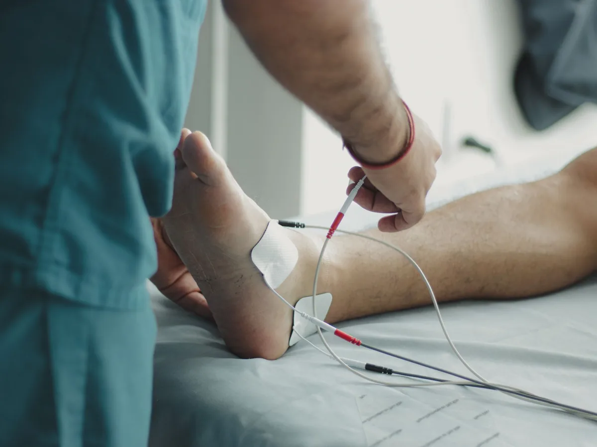
The nerve conduction study is the first half. Small sticky electrodes go on the skin over a nerve and the muscle it supplies. A brief electrical pulse stimulates the nerve at one point, and the machine records what arrives at the other, timing the journey and measuring the size of the signal. Distance divided by travel time is the conduction velocity.
The EMG is the second half, and the part people worry about. A fine needle, thinner than the one used to draw blood, is placed in a few muscles. It is not injecting anything; it is listening. The doctor watches and hears the electrical activity of the muscle at rest and while you gently tense it, which shows whether a weak muscle is losing its nerve supply and roughly how long that has been going on.
The needle test tells you about muscles and the nerves running to them. Not every evaluation needs it: for typical numbness in both feet with no weakness, the nerve conduction study often answers the question on its own.
Who orders them, and who does them
A primary care clinician, neurologist, physiatrist, podiatrist or surgeon can order the tests. They are usually performed in a neurology or rehabilitation department. The American Association of Neuromuscular and Electrodiagnostic Medicine advises that a suitably trained doctor should perform all needle EMG testing, while a trained technologist may perform nerve conduction studies under a doctor’s supervision.
For most people with diabetes and the usual pattern of numbness that creeps up from the toes, these tests are not needed at all. The American Diabetes Association’s 2026 Standards of Care say electrophysiological testing or referral to a neurologist is rarely needed, except when the picture is atypical — sudden, asymmetric, not length-dependent, or involving weakness — or when the diagnosis is unclear. Which clinician does what.
How to prepare
The AANEM asks patients not to use body lotion on the day of the test. Oils and creams get between the electrodes and your skin.
There is no fasting and no sedation, so normal activities before and after are fine.
Aspirin and blood thinners such as warfarin matter for the needle part. In practice, studies have found needle EMG to be safe without stopping anticoagulants, but the decision belongs to the doctor doing the test.
Tell them about a pacemaker or defibrillator. Modern devices are not a barrier to the test, but the team will take that into account, and nerve conduction studies are avoided in people with intracardiac catheters or external pacing wires.
Cold limbs slow nerve conduction, and a cold foot can look like a nerve problem. Testing labs warm the limb to a set temperature for that reason. Wear socks to the appointment in winter, and say if your feet are cold.
What it feels like, and how long it takes
The whole appointment usually takes 20 to 90 minutes, depending on how many nerves are studied. The nerve conduction part feels like quick, mild shocks, described by the AANEM as a brief tingling sensation. It is startling rather than painful. The needle part can hurt a little, and some people find the muscles feel bruised for a day afterward. Neither test has lasting side effects, and you can do your normal activities as soon as you leave.
If you are anxious about the needle, say so before you start. Fewer muscles can sometimes be tested, and knowing what is happening helps more than most people expect.
Reading the result
You will usually be given a report rather than a diagnosis. Two numbers carry most of the meaning.
| What changed | What it usually means |
|---|---|
| Slow conduction velocity, long latency | The insulation (myelin) around the nerve is damaged. This is the demyelinating pattern, seen in some inherited and immune neuropathies, and where a nerve is compressed. |
| Small signal (amplitude), speed nearly normal | Fewer nerve fibers are working. This is the axonal pattern, and it is what most neuropathies from diabetes, alcohol, B12 deficiency or chemotherapy look like. |
| Changes only across one point on a nerve | The nerve is being compressed there, for example at the fibular head at the outside of the knee. That is a common cause of foot drop. |
| A pattern that follows a nerve root rather than a nerve | The problem is likely in the spine rather than in the leg. Telling a back problem from neuropathy. |
| Fibrillations and other changes on the needle test | A muscle has lost part of its nerve supply. The pattern also indicates roughly how recent that loss is. |
The report is a description of the nerves, not the cause. Finding the cause still takes the history, the examination and blood tests. The tests that find the common causes.
What a normal result means
It means the large, fast, heavily insulated fibers are working. Those carry vibration, position sense and muscle signals. The small fibers that carry burning pain and temperature are thin or bare, conduct slowly, and are not measured by a standard nerve conduction study at all.
So a person can have severe burning in both feet, a confidently normal nerve conduction study and EMG, and a real neuropathy. This is one of the most common sources of frustration we hear about. Small fiber neuropathy is the explanation.
When small fiber neuropathy is suspected, the usual next step is a skin biopsy: a 3 mm punch of skin from the lower leg, in which the nerve fiber endings are counted under a microscope. A European Federation of Neurological Societies and Peripheral Nerve Society task force rated distal leg skin biopsy with nerve fiber counting as a reliable technique for diagnosing small fiber neuropathy, its strongest level of recommendation. Autonomic tests, such as a sweat test, can add to the picture.
Normal results are also useful in their own right. They make an immune neuropathy, a nerve-root problem or a compressed nerve much less likely, which changes what is worth looking for next.
Common questions
Does a nerve conduction study hurt?
The shocks are brief and mild, more startling than painful. The needle part of the EMG can hurt a little while the needle is being moved, and the muscle may feel bruised for a day. There is no lasting soreness and no sedation is needed.
How long does a nerve conduction study take?
Usually 20 to 90 minutes for the whole appointment, including the needle EMG if it is done. A study of one nerve is much quicker than a survey of both legs.
Can a nerve conduction study be normal if I have neuropathy?
Yes, and often is if the problem is in the small fibers. Standard testing measures large fibers only. Burning pain, temperature changes and pinprick loss with a normal study point to small fiber neuropathy, which is diagnosed by examination and, where needed, a skin biopsy.
Do I need to stop my blood thinner?
Usually not. Needle EMG has been found safe in people taking anticoagulants and antiplatelet medicines, but tell the doctor doing the test what you take and let them decide. Never stop a blood thinner on your own.
What is the difference between a nerve conduction study and an EMG?
The nerve conduction study measures signals traveling along nerves using surface electrodes. The EMG measures the electrical activity of muscles using a fine needle. They answer different questions and are usually done together.
Related reading
Small fiber neuropathy · The 10-gram monofilament test · Which doctor treats neuropathy · Back nerve pain versus neuropathy · Numb feet · Foot drop
Sources and further reading: American Association of Neuromuscular and Electrodiagnostic Medicine, FAQs before electrodiagnostic testing and types of tests, on duration, preparation, sensations and who performs the tests. Ramani PK, Lui F, Arya K, Nerve conduction studies and electromyography, StatPearls, 2025, on large-fiber measurement, limb temperature, axonal and demyelinating patterns, anticoagulation and implanted devices. NINDS, peripheral neuropathy, on what each test measures and on neurodiagnostic skin biopsy. American Diabetes Association Standards of Care in Diabetes—2026, section 12, on when electrophysiological testing is needed. Lauria G et al., EFNS/PNS guideline on the use of skin biopsy in the diagnosis of small fiber neuropathy, Eur J Neurol 2010;17:903–912.
This page is general education and is not a diagnosis or a treatment plan. Clinical review by Dr. Biernacki is pending; the review date will be shown here once complete.
For our Michigan neighbors
Told your nerve test
was normal?
A normal study does not mean nothing is wrong, and the next step depends on the pattern in your feet. Dr. Biernacki’s Michigan practice can take a look.
Everywhere else, take this page to your own clinician.

Dr. Tom Biernacki, DPM, FACFAS is a double board-certified foot and ankle surgeon (ABFAS and ABPM) and a Fellow of the American College of Foot & Ankle Surgeons. He is the founder of Balance Foot & Ankle in Howell and Bloomfield Township, Michigan, where peripheral neuropathy, diabetic foot risk and loss of protective sensation are part of his weekly clinic. His patient education videos have drawn more than 950,000 subscribers on YouTube. Managing Neuropathy is written by its editorial team under his direction, to put the explanation he gives in the exam room in front of the people who cannot get to one — including when a product is worth buying and when it is not.