Treatment
Foot drop exercises: a safe home routine, and a quick self-check
Exercise can’t regrow a nerve. It can keep the ankle from stiffening, strengthen what still works, and keep you steady on your feet while the cause is found and treated.
Written by the Managing Neuropathy editorial team under the direction of Dr. Tom Biernacki, DPM, FACFAS · Clinical review pending · Last updated 26 September 2026
The short answer. Get a new foot drop examined first. Then a home routine has four jobs: stretch the calf and Achilles daily so the ankle doesn’t stiffen, work the muscles that lift the foot and toes at whatever level you can, train your balance, and practice walking safely, usually in a brace. Exercise can’t regrow a nerve, so recovery depends on the cause. Wear shoes, hold on to something steady, and check numb skin afterward.
What exercise can and can’t do
What it can do. When the muscles that lift the foot are weak, the calf works unopposed. Over time the Achilles tendon can shorten and the ankle stiffen with the foot pointing down. A stiff ankle is harder to brace, and it can block a normal step even if the nerve recovers. Daily stretching is the standard defense. Exercise also maintains the muscles that still have nerve supply, and it trains steadier walking.
What it can’t do. It can’t regrow a nerve, and a muscle with no nerve supply at all can’t be strengthened. Recovery depends on the cause and the level. Pressure on the peroneal nerve at the outside of the knee often improves once the pressure stops, while foot drop from a progressive disease is usually permanent. That is why finding the cause and the level comes first. Peroneal neuropathy, a common cause, is covered here.
Check yourself: a simple foot drop test
Do each check on both sides and compare.
- Seated foot lift. Feet flat and heels down, lift the toes and the front of each foot as high as you can.
- Heel walk. Holding the counter, take a few steps on your heels. A weak foot drops or slaps down. Skip this if you feel unsteady.
- Turning-in check. Sitting, turn the sole inward against gentle pressure from your hand, then outward. Turning outward is often weak with foot drop. If turning inward is weak too, the problem probably involves more than the peroneal nerve at the knee, such as a nerve root in the lower back.
What it can’t tell you. These checks don’t find the cause, and they can look normal when partial weakness shows only as the toe catching late in the day. A clinician watches you walk, grades strength from 0 (no movement) to 5 (normal), and may order nerve conduction studies and electromyography (EMG) or imaging.
Before you start: safety for numb feet
Many causes of foot drop also dull feeling in the foot, and numb skin can’t warn you about a rub or a blister.
- Keep shoes on for standing exercises and walking practice, even indoors, and a sock or shoe under any band.
- Check your skin afterward, including anywhere a brace or strap presses. The daily foot check is here.
- Stop for pain or a wound, and get a wound seen before you carry on.
Goal 1: keep the calf and Achilles from tightening
Do this every day, from the start, unless your surgeon or therapist has said otherwise. Stretch within a comfortable range, and don’t force it into pain.
- Towel stretch, seated. With the leg straight, loop a towel around the ball of the foot and pull gently until you feel the calf stretch. Hold 30 seconds, relax, and repeat.
- Standing calf stretches. Facing a counter, step the affected leg back with the heel down, toes forward and knee straight, and bend the front knee. Then take a shorter step and bend both knees, which stretches the soleus, the second calf muscle. Hold each 30 seconds, three times.
A night splint holds the ankle near a right angle while you sleep. The evidence for splinting alone is thin: two small trials in Charcot-Marie-Tooth disease, an inherited neuropathy, found night splints no better than none for ankle flexibility. Use one if your clinician advises it, alongside the stretching, and check the skin under the straps each morning.
Goal 2: strengthen the muscles that lift and turn the foot
Muscles at the front of the shin lift the foot and toes, a movement called dorsiflexion. The peroneal muscles on the outer shin turn the foot outward, called eversion. A peroneal nerve problem can weaken both. Start at the level you can manage. For lifting, repetition is the key.
Sitting, loop a belt or towel under the ball of the foot and hold both ends. Try to lift the foot, and pull just enough to help it up. Guide it outward with your hand the same way. With no movement yet, this keeps the joint moving. Tell your therapist about the first flicker of movement.
Heel down, lift the front of the foot toward your shin, then lower it slowly. An NHS Tayside (Scotland) exercise sheet for drop foot, written for people with multiple sclerosis, suggests 20 to 30 lifts, two or three sets, if you can. Add toe lifts, and lift the outer edge of the foot.
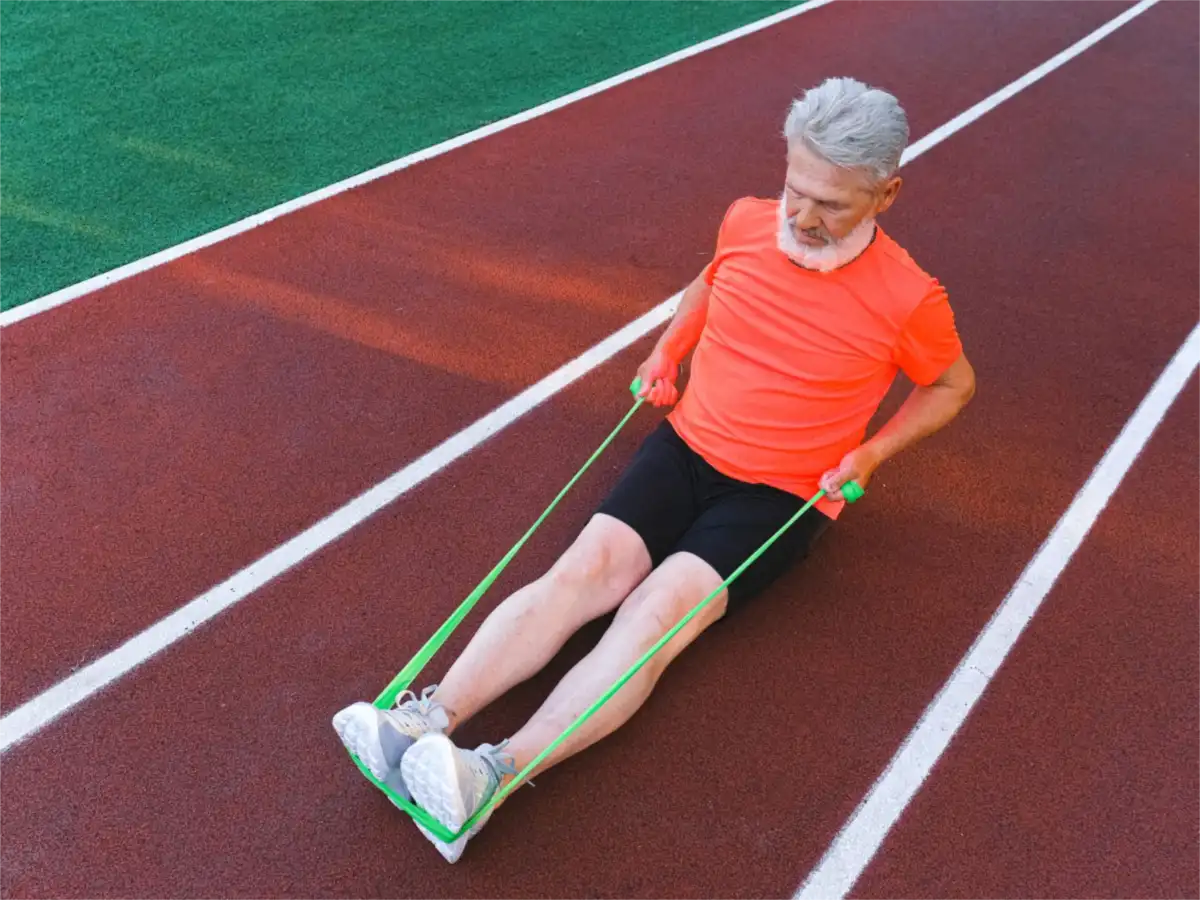
Lift against your other foot resting on top, or against a band anchored to something solid. The same program suggests 10 to 20 repetitions, three sets. For eversion, anchor the band on the inner side and turn the foot out against it, as your therapist directs.
Goal 3: balance and fall safety
Foot drop raises the risk of falls. At the counter, balance on one leg for up to 30 seconds, holding on as much as you need, three to five times on each side. More balance moves are in our neuropathy exercise routine. At home, the NHS advises clear floors, no loose rugs or trailing cables, good lighting, stair handrails, and a walking aid if you need one. If the brace comes off at night, add a night-light.
Goal 4: walking practice, with and without the brace
With the brace. An ankle-foot orthosis, or AFO, holds the foot near a right angle so the toe clears the ground. It prevents trips, though it doesn’t treat the nerve. Practice on flat floors first, then curbs and stairs, using a rail. Build up wearing time over days, and check your skin each time it comes off. Choosing and fitting a foot drop brace.
Without the brace. Only if your therapist adds it as strength returns: short walks indoors, with a counter or rail within reach. If the toe starts catching, stop for the day.
Beyond exercise: electrical stimulation and surgery
Functional electrical stimulation (FES) uses a cuff below the knee, or an implant, to stimulate the peroneal nerve with each step so the foot lifts. It needs a working nerve between the spinal cord and the muscle, so it suits foot drop that starts in the brain or spinal cord, as after a stroke or with multiple sclerosis, and usually not a damaged nerve in the leg. The National Institute for Health and Care Excellence (NICE), which advises the NHS, supported its use for these causes in 2009. In a trial of 197 stroke survivors, walking speed improved over 30 weeks with either FES or an AFO, with no significant difference between them, and the FES group was more satisfied. Electrodes can irritate the skin, which matters where it is numb.
Surgery. A pinched peroneal nerve that is getting worse quickly, or hasn’t improved after about three months, may be released surgically. Timing matters, because a muscle without its nerve for 18 months is unlikely to recover much function. For long-standing foot drop, a tendon transfer reroutes a working tendon, most often the posterior tibial tendon, to lift the foot. The American Orthopaedic Foot & Ankle Society calls it reasonable to consider when no function has returned by about a year. A very stiff ankle may also need the Achilles lengthened, one more reason to stretch now. When the cause is in the back, a spine specialist decides. Back nerve pain versus neuropathy.
When to get help fast
- Call 911 for foot drop that comes on suddenly, especially with face or arm weakness, trouble speaking or loss of balance. These are stroke signs.
- Emergency care for numbness in the groin or buttocks, or new bladder or bowel trouble, especially with back pain. These can mean cauda equina syndrome, pressure on the nerve roots at the base of the spine, which is a surgical emergency.
- Emergency care for weakness in both feet that comes on over hours or days, or weakness spreading upward, as in Guillain-Barré syndrome. Call 911 if breathing becomes difficult.
- Call your doctor right away for foot drop that appears after a leg injury or an operation, or while you are in a cast or a tight bandage. Go to an emergency room if the leg is also very painful, swollen or tight, which can mean compartment syndrome, a surgical emergency.
- Within days for foot drop that is getting worse.
- Stop the routine and call for new pain, a wound or a fall.
Common questions
Can foot drop be fixed with exercises?
Not by exercise alone. Exercise can’t regrow a nerve, and anything still pressing on the nerve has to be removed first. What exercise does is keep the ankle flexible, maintain the muscles that still work and make walking safer, so the ankle is ready if the nerve recovers.
How do you test for foot drop at home?
Sitting with your heels down, lift the front of each foot. Then, holding a counter, take a few steps on your heels. A foot that won’t lift, or slaps down, suggests weakness, and a weak turn inward hints at a problem higher up, such as the lower back. None of this finds the cause. That needs an examination.
How often should you do foot drop exercises?
Stretch the calf every day. Strengthening programs vary: an NHS Tayside exercise sheet for drop foot suggests 20 to 30 lifts for two or three sets, and a general ankle program from the American Academy of Orthopaedic Surgeons lists band work three days a week. Let your physical therapist set your amount, stop for pain, and ease off if the foot tires.
Should I walk without my foot drop brace to build strength?
Only if your therapist suggests it. The weakness comes from the nerve, wearing the brace is unlikely to add to it, and walking without one raises the risk of tripping. Your therapist may add short brace-free practice as strength returns. Otherwise, wear the brace and do the strengthening exercises separately.
How long does foot drop take to recover?
It depends on the cause. A mild pressure injury with the nerve fibers intact usually recovers within about three months. Where fibers were lost, recovery can take up to a year. Foot drop from a progressive condition is usually permanent. If a pressure-related drop isn’t improving by three months, ask about nerve testing and a surgical opinion.
Related reading
Foot drop: finding the level · Choosing a foot drop brace · Peroneal neuropathy · Neuropathy exercises for the legs and feet · Nerve conduction study and EMG · The daily foot check
Sources and further reading: NHS Tayside, Managing drop foot, written for people with MS. AAOS OrthoInfo, Foot and ankle conditioning program. Nori SL, Stretanski MF, Foot drop, StatPearls, 2025. Lezak B et al., Peroneal nerve injury, StatPearls, 2024. Baima J, Krivickas L, Evaluation and treatment of peroneal neuropathy, Current Reviews in Musculoskeletal Medicine, 2008. Carolus AE et al., The interdisciplinary management of foot drop, Deutsches Ärzteblatt International, 2019. Rose KJ et al., Interventions for increasing ankle range of motion in patients with neuromuscular disease, Cochrane Review, 2022. NICE, Functional electrical stimulation for drop foot of central neurological origin, 2009. Kluding PM et al., Foot drop stimulation versus ankle foot orthosis after stroke: 30-week outcomes, Stroke, 2013. Cleveland Clinic, functional electrical stimulation. American Orthopaedic Foot & Ankle Society, Foot drop treatment (tendon transfer surgery). Mayo Clinic, foot drop diagnosis and treatment, 2025. NHS, foot drop, 2025. Warning signs: CDC, stroke; AAOS OrthoInfo, cauda equina syndrome; NINDS, Guillain-Barré syndrome. NIDDK, diabetes and foot problems.
This page is general education and is not a diagnosis, a treatment plan, or a substitute for an exercise program designed for you. Clinical review by Dr. Biernacki is pending; the review date will be shown here once complete.
For our Michigan neighbors
Toe still catching
despite the exercises?
A foot drop that isn’t improving needs its level found, and a braced foot with numb skin needs watching. Dr. Biernacki’s Michigan practice can do both.
Everywhere else, take this page to your own clinician.

Dr. Tom Biernacki, DPM, FACFAS is a double board-certified foot and ankle surgeon (ABFAS and ABPM) and a Fellow of the American College of Foot & Ankle Surgeons. He is the founder of Balance Foot & Ankle in Howell and Bloomfield Township, Michigan, where peripheral neuropathy, diabetic foot risk and loss of protective sensation are part of his weekly clinic. His patient education videos have drawn more than 950,000 subscribers on YouTube. Managing Neuropathy is written by its editorial team under his direction, to put the explanation he gives in the exam room in front of the people who cannot get to one — including when a product is worth buying and when it is not.